Wednesday, February 10, 2010

Wednesday, February 3, 2010
CHEM 1311
Alrighty… Time to dive right in with some awesome websites that will help with information on the first two chapters we will be tested on…
First, a few really great online calculators to help check your homework when converting back and forth between different units of measure in the decimal system. I recommend just using these to check your answers so that you are working the problems the correct way…after all, we won’t have these for help on the exams… :(
http://www.worldwidemetric.com/metcal.htm
http://www.unit-conversion.info/metric.html
Next, I’ve got an EXCELLENT site on converting to numbers to Scientific Notation. This site explains the rules for addition/subtraction and multiplication and division, AND (this is the best part!) offers an interactive quiz for you to practice. 30 minutes with this site, and you’re golden.
http://janus.astro.umd.edu/astro/scinote/help.html
And…the moment you’ve all been waiting for…a calculator to check your answers for Polyatomic Nomenclature conversions!!! YES!! Courtesy of UNC – Chapel Hill, you can put together all kinds of combinations. Plus, there’s a plethora of other great chemistry information and tutorials here. Basically a JACKPOT!! :)
http://www.shodor.org/unchem/basic/nomen/polycalc.html
Between these 4 sites, I think just about everything we could possibly be asked on the exam is covered in some form or another. But, later this week, I’ll post all my notes that I’ve taken from the book on Chapters 1 and 2. Look for those probably sometime on Saturday.
Happy Studying!
-L
Monday, January 18, 2010
Officially Changed My Major…I’ve Become Good At That Over The Years
Classes start the day after tomorrow…although since it’s 2:15 am right now, I guess I can say that they start tomorrow, instead. And, in true “LB” fashion, I officially changed my major tonight. Not that it’s really different from what I had discussed with anyone last semester… I started off by declaring a Nursing major. But, I decided midway through the Anatomy class that I wanted to go completely crazy (yes, I need to have my head examined!) and apply to Medical School. And, tonight, I found out the Austin Community College offers a Pre-Medical Associate Degree. It has all the classes I need as prerequisites to apply to medical school, plus a few extra liberal arts electives that I would need to fulfill if I decided to pull the plug on spending the rest of my life in medical school and get my BSN at University of Texas, instead. And, I figure, hey…if I’m doing all this work, anyway, shouldn’t I get some kind of a nice diploma out of the deal? I say, “YES!” :) Anyway, I’ve got several of the electives finished, and anything that is not directly related to the medical school requirements, I will wait until after I’ve taken the MCAT and applied to schools to complete. As I was thinking about it before, I realized that I would be sitting around doing absolutely nothing for about a year once I complete the hardcore prerequisites, anyway, so the extra classes will give me something to do while I wait to find out if anyone will take me (PLEASE! SOMEONE TAKE ME!!), and will also get me prepared to transfer into UT if they decide not to take me. Either way, I think it’s a good, solid plan, and I feel very at peace with it.
So…on-tap for this semester is my heaviest course load yet. I’m taking 11 hours while working full time (yes…I’m on crack). The good news is that the text for my Cellular and Molecular Biology class is the same one that we used for AP Biology when I was in high school…only this sucker is 5 editions NEWER!! Wow…I feel very old. Anyway, my point, here, is that the material will be more of a review, so I’m very glad for that. I’m also taking General Chemistry 1. I took this class at University of Colorado, but it was about 15 years ago, and I barely remember a thing. So…this will be review as well. AWESOME! And, finally, there will be US History II. This is one of those elective classes that will be required for the UT BSN as well as the Pre-Med AD. And because I’m taking mostly review stuff this semester, I figured it would be a good time to max out my course load… I take that back… I don’t think any time is a “good” time to max out my course load, but if it has to be done, this would be the time for it. After this semester, there really won’t be any more “eeek” room until after I take the MCAT and start applying to schools at the end of NEXT Spring semester.
Anyway, here’s wishing everyone a great semester with good professors and easy tests!! A girl can dream, right?!??? ;)
Friday, December 11, 2009
It’s the moment of Truth…
Last night for studying, kids… One last practical to take, and this semester is in the books. I think I probably need a 30 out of 40 to keep an A in the class. This is a guess, though, since I don’t know what I got on the last exam. I don’t think the last exam was as air-tight as I’ve been on previous ones… I wrote 11 pages for the essays, and that wasn’t counting the extra credit diagram. But, I kept thinking of things I could have added once I left. Of course, after literally THREE HOURS of non-stop writing, I HAD TO finish. I really needed to go to the “little girls’ room” at the end of hour 2, but since I was nowhere near finished and they won’t let you leave, I had to stick it out for as long as all those little sphincters would let me (haha…I’m hilarious…or just sharing TMI…but it’s true, and THAT, my friends, is EXAM MATERIAL you can take to the bank!!).
Anyway, good luck to everyone with your studying tonight. I think that my last post had a link to just about everything, and if not, it was pretty darn close. It’s been a GREAT semester, and despite all the long nights and stress from cramming all this good information into my head, I really enjoyed it and had fun meeting all of you! I will be keeping this blog around for my future classes since it seems to have been a pretty decent study tool. So…if we have more classes together, keep this page bookmarked! And if we don’t have more classes together and you just want to see me get my mind MELDED when I take O-Chem in the fall, stick around for that, too! It’s going to be GOOD TIMES!! Next up, though: Cellular Biology and General Chemistry 1. I had chem 1 and some cell bio in college, but good lord, that was almost 15 years ago that I took those classes, and I can barely remember a darn thing!! (I can’t believe I just put that number in PRINT!)
So, good luck in your future studies, and hopefully, I’ll be seein’ ya somewhere again in the future!! :)
-L
Thursday, December 10, 2009
A Little Help For the Last Practical! WoooHooo!! :)
Found a great website thanks to my alma mater... Wish I'd found this earlier in the year... It's actually pretty good. This will give you a set of links to models for the urinary system and the reproductive system (male and female):
Go Buffs!
http://www.colorado.edu/intphys/iphy3415/models/
Histology:
http://www.kumc.edu/instruction/medicine/anatomy/histoweb/
Digestive Lab
Digestive Models:
http://nhscience.lonestar.edu/bioL/ap2model/digestive.htm
Digestive Slides:
Salivary Glands:
http://nhscience.lonestar.edu/bioL/digestive/salivary.htm
http://www.austincc.edu/histologyhelp/organs/op_salivary.html
Esophagus Slides:
http://nhscience.lonestar.edu/bioL/digestive/esophagu.htm
http://www.austincc.edu/histologyhelp/organs/oq_esophagus.html
Stomach Slides:
http://nhscience.lonestar.edu/bioL/digestive/stomach2.htm
http://nhscience.lonestar.edu/bioL/digestive/stomach.htm
http://www.austincc.edu/histologyhelp/organs/or_stomach.html
Small Intestine:
Duodenum : http://nhscience.lonestar.edu/bioL/digestive/smintest.htm
Duodenum: http://www.austincc.edu/histologyhelp/organs/os_duodenum.html
Jujunum: http://nhscience.lonestar.edu/bioL/digestive/jejunum.htm
Ilium: http://nhscience.lonestar.edu/bioL/digestive/ileum.htm
Ilium: http://www.austincc.edu/histologyhelp/organs/ot_ileum.html
Pancreas: http://district.bluegrass.kctcs.edu/shirley.whitescarver/BIO139Lab/BIO139/139Lab12/Lab12EndocrineSlides.html (scroll to the bottom of the page)
Liver:
http://www.austincc.edu/histologyhelp/organs/ov_liver.html
http://www.kumc.edu/instruction/medicine/anatomy/histoweb/gitract/gitract.htm
Urinary System Lab:
Kidney: http://www.austincc.edu/histologyhelp/organs/ow_kidney.html
Kidney - Nephron: http://nhscience.lonestar.edu/bioL/urinary/nephron.htm
Bladder Slides:
http://nhscience.lonestar.edu/bioL/urinary/bladder3.htm
http://nhscience.lonestar.edu/bioL/urinary/bladder.htm
Kideny Model:
http://www.colorado.edu/intphys/iphy3415/models/kidney.html
Reproductive Systems Lab:
Testis Slide: http://www.austincc.edu/histologyhelp/organs/oy_testis.html
Penis Crossection: http://nhscience.lonestar.edu/bioL/reproductive/penis.htm
Male Reproductive Models:
http://www.colorado.edu/intphys/iphy3415/models/repro_male.html
http://www.colorado.edu/intphys/iphy3415/models/male_2005.html
Female Reproductive Models:
http://www.colorado.edu/intphys/iphy3415/models/repro_female.html
http://www.colorado.edu/intphys/iphy3415/models/female_2005.html
Go Buffs!
http://www.colorado.edu/intphys/iphy3415/models/
Histology:
http://www.kumc.edu/instruction/medicine/anatomy/histoweb/
Digestive Lab
Digestive Models:
http://nhscience.lonestar.edu/bioL/ap2model/digestive.htm
Digestive Slides:
Salivary Glands:
http://nhscience.lonestar.edu/bioL/digestive/salivary.htm
http://www.austincc.edu/histologyhelp/organs/op_salivary.html
Esophagus Slides:
http://nhscience.lonestar.edu/bioL/digestive/esophagu.htm
http://www.austincc.edu/histologyhelp/organs/oq_esophagus.html
Stomach Slides:
http://nhscience.lonestar.edu/bioL/digestive/stomach2.htm
http://nhscience.lonestar.edu/bioL/digestive/stomach.htm
http://www.austincc.edu/histologyhelp/organs/or_stomach.html
Small Intestine:
Duodenum : http://nhscience.lonestar.edu/bioL/digestive/smintest.htm
Duodenum: http://www.austincc.edu/histologyhelp/organs/os_duodenum.html
Jujunum: http://nhscience.lonestar.edu/bioL/digestive/jejunum.htm
Ilium: http://nhscience.lonestar.edu/bioL/digestive/ileum.htm
Ilium: http://www.austincc.edu/histologyhelp/organs/ot_ileum.html
Pancreas: http://district.bluegrass.kctcs.edu/shirley.whitescarver/BIO139Lab/BIO139/139Lab12/Lab12EndocrineSlides.html (scroll to the bottom of the page)
Liver:
http://www.austincc.edu/histologyhelp/organs/ov_liver.html
http://www.kumc.edu/instruction/medicine/anatomy/histoweb/gitract/gitract.htm
Urinary System Lab:
Kidney: http://www.austincc.edu/histologyhelp/organs/ow_kidney.html
Kidney - Nephron: http://nhscience.lonestar.edu/bioL/urinary/nephron.htm
Bladder Slides:
http://nhscience.lonestar.edu/bioL/urinary/bladder3.htm
http://nhscience.lonestar.edu/bioL/urinary/bladder.htm
Kideny Model:
http://www.colorado.edu/intphys/iphy3415/models/kidney.html
Reproductive Systems Lab:
Testis Slide: http://www.austincc.edu/histologyhelp/organs/oy_testis.html
Penis Crossection: http://nhscience.lonestar.edu/bioL/reproductive/penis.htm
Male Reproductive Models:
http://www.colorado.edu/intphys/iphy3415/models/repro_male.html
http://www.colorado.edu/intphys/iphy3415/models/male_2005.html
Female Reproductive Models:
http://www.colorado.edu/intphys/iphy3415/models/repro_female.html
http://www.colorado.edu/intphys/iphy3415/models/female_2005.html
Sunday, November 29, 2009
Flash Cards for Unit 4 Exam
Hey All-
Hope you have all had a great Thanksgiving. I'm putting together a deck of flashcards on quizlet.com for the last 4 chapters of material (I'll be using them as my 4th take-home assignment). But, I'll be posting links to the different chapters as I finish. For the Digestive System Chapter (Chapter 22), here is the first link:
Digestion
or the full URL is http://quizlet.com/1490330/digestion-flash-cards/
I am still adding to these, obviously, but I've got the minimum 20 cards required for full credit on the assignment as of now, which is why I'm posting. Be sure to check back as I continue to update the material and add more chapters tomorrow and throughout the week!
Happy Studying!! :)
-L
Hope you have all had a great Thanksgiving. I'm putting together a deck of flashcards on quizlet.com for the last 4 chapters of material (I'll be using them as my 4th take-home assignment). But, I'll be posting links to the different chapters as I finish. For the Digestive System Chapter (Chapter 22), here is the first link:
Digestion
or the full URL is http://quizlet.com/1490330/digestion-flash-cards/
I am still adding to these, obviously, but I've got the minimum 20 cards required for full credit on the assignment as of now, which is why I'm posting. Be sure to check back as I continue to update the material and add more chapters tomorrow and throughout the week!
Happy Studying!! :)
-L
Thursday, October 29, 2009
Excellent Websites for Studying for the Practical
I'm missing some areas in here (the human brain model, the sheep brain, brain slides, human eye, cow eye, retina slide, human ear, cochlear cross-section cochlear slide, olfactory epithelium slide, and tongue slide), but I have SEEN most of these on the web in my search for other things, and I will be working on this tomorrow, and updating this post throughout the day. :)
-L
I decided to Ask Google how to study for Practical #3, and wouldn't you know...Google has a PhD in Anatomy! Below, I've got a list of EXCELLENT websites to help study for each of the different labs, section by section. Good stuff!! :) And, what's really awesome, is that a lot of the sites have everything labeled and give some explanation into what you are looking at. Happy Studying!
Complete JACKPOT:
http://bioweb.uwlax.edu/aplab/Table_of_Contents/table_of_contents.html
First I found a single page with a good slide of a motor neuron. But, when I looked at the URL, I realized there was a whole INDEX of pages that are designed for students in the 2304 class to help with histology. Wish I'd found this sucker when we did the big histology practical! BOO!
Anyhow, the main page is:
http://www.austincc.edu/histologyhelp/
Then from there, you can navigate to tissues:
http://www.austincc.edu/histologyhelp/tissues/index.html
and then organs, which is where you will find our endocrine slides:
http://www.austincc.edu/histologyhelp/organs/index.html
And an awesome, all-purpose neurology jagon website:
http://mindsci-clinic.com/neuro_jargon.htm
Nervous System Lab
Neuron Model:
Cell body, nucleus, chromatophilic bodies, dendrite, telodendria, axon, axon terminal, axon hillock, synapse, Schwann cells, myelin sheath, & nodes of Ranvier
http://bioweb.uwlax.edu/aplab/Table_of_Contents/Lab_06/Neuron_Model_1/neuron_model_1.html
http://bioweb.uwlax.edu/aplab/Table_of_Contents/Lab_06/Neuron_Model_1/Neuron_Model_1a/neuron_model_1a.html
http://nhscience.lonestar.edu/bioL/ap1model/labeled/neuron.jpg
Motor Neuron Slide:
Cell body, cellular processes
Hey, hey! What do you know... a site from our school!
http://www.austincc.edu/histologyhelp/tissues/tx_nerv_tis.html
Peripheral Nerve Slide:
Nerve fibers, fascicles, epineurium, perineurium
http://bioweb.uwlax.edu/aplab/Table_of_Contents/Lab_06/Nerve_1/Nerve_1a/nerve_1a.html
http://ect.downstate.edu/courseware/neurohisto_intro/slide8.html
Spinal Cord Models:
Gray horns, (anterior, lateral and posterior), White Columns (Anterior lateral and Posterior), Gray and White Commissures, Anterior medial fissure, Dorsal medial sulcus Meninges (dura mater, arachnoid, pia mater), Subarachnoid space, Dorsal root,Dorsal root gangilion,Ventral root, Epidural space, Rami, (dorsal ramus, ventral ramus, gray ramus communicantes)
Rockin' Spinal Cord Models that allow you to quiz yourself with labeled and non-labeled pictures!
http://classroom.sdmesa.edu/anatomy/ModelPages/spinal_cord.htm
Also, I had trouble finding a model with the White Commissure listed. But, here is a cross section that shows it, and a link to Wikipedia that explains:
http://biology.clc.uc.edu/Fankhauser/Labs/Anatomy_&_Physiology/A&P202/CNS_Histology/Spinal_Cord/sp_cd_jpgs/Spinal_Cord_PC271498_lbd.JPG
http://en.wikipedia.org/wiki/Anterior_white_commissure
Had trouble finding the Rami, but this drawing diagrams very well:
http://instruct.uwo.ca/anatomy/530/segm.gif
Also:
http://images.google.com/imgres?imgurl=http://upload.wikimedia.org/wikipedia/commons/thumb/e/ee/Gray799.svg/250px-Gray799.svg.png&imgrefurl=http://en.wikipedia.org/wiki/Gray_ramus_communicans&usg=__qUXCvvpEp5jq-MU-9iTXPLFa0f0=&h=222&w=250&sz=31&hl=en&start=8&sig2=C5MPFfY1uQFQtlrMe7Nwww&um=1&tbnid=9BHh2mc5_tWNJM:&tbnh=99&tbnw=111&prev=/images%3Fq%3Dgray%2Brami%2Bcommunicans%2Bmodel%26hl%3Den%26um%3D1&ei=AFXqSortMMSztgfR-9w_
Spinal Cord Cross Section:
Gray horns (anterior, lateral posterior), white columns (anterior, lateral, posterior), central canal
http://nhscience.lonestar.edu/bioL/ap1model/labeled/spinalcord3.jpg
http://bioweb.uwlax.edu/Aplab/Table_of_Contents/Lab_07/Spinal_Model_1/spinal_model_1.html
http://bioweb.uwlax.edu/Aplab/Table_of_Contents/Lab_07/Spinal_Model_2/spinal_model_2.html
Nerve and Nerve Plexi / Cranial Nerves:
Using appropriate models, be able to identify all 12 pairs of cranial nerves as well as the primary nerves of the cervical plexus, brachial plexus, lumbar plexus, and sacral plexus. Be sure to review the function of each.
http://nhscience.lonestar.edu/bioL/ap1model/labeled/spinalcord.jpg
The secret to remembering these is to know that the are numbered in order from rostral to caudal end, starting with the Olfactory nerve. From there, you can count off the nerves and use the mneumonic "OLd OPie OCcassionally TROmps TRIGonometry And Feels VEry GLOomy, VAGUe And HYPOactive."
http://content.answers.com/main/content/img/oxford/Oxford_Body/019852403x.cranial-nerves.1.jpg
http://academic.kellogg.edu/herbrandsonc/bio201_McKinley/f15-24l_cranial_nerves_c.jpg
Quiz yourself on Cranial Nerves!
http://www.dontbeasalmon.net/elearning/term3spotters/docs/qa/CN.html
Sympathetic Efferent Pathway Models:
Paravertebral sympathetic ganglia, Prevertebral sympathetic ganglia
The paravertebral sympathetic ganglia are also known as the sympathetic trunk ganglia or chain ganglia. The prevertebral ganglia are located further away from the sympathetic trunk (more distal?), and are also known in our text as collateral ganglia.
http://www.ncbi.nlm.nih.gov/bookshelf/br.fcgi?book=neurosci&part=A1391&rendertype=figure&id=A1393
Human Brain Model:
Cerebrum:
Cerebral hemispheres (left and right), transverse fissure, longitudinal fissure, lateral sulcus, central sulcus, parieto-occipital sulcus, precentral gyrus, postcentral gyrus, frontal lobe, parietal lobe, temporal lobe, occipital lobe, insula, basal nuclei, corpus callosum, septum pellucidum, fornix, cortex, internal capsule, olfactory bulbs, olfactory tracts, optic nerves, optic chiasma, optic tracts, roots of nerves III – XII, lateral ventricles, choroid plexus
http://lh4.ggpht.com/_lRGislJejZ0/SRp7sb6QjHI/AAAAAAAAB8c/xeGXGZP0LUY/image20_thumb4.png?imgmax=800
http://mindsci-clinic.com/cerebral_lobes_and_insula.htm
Quiz yourself on the internal structures:
http://www.dontbeasalmon.net/elearning/term3spotters/docs/qa/index.html
Diencephalon:
Pituitary gland, mammillary bodies, thalamus, massa intermedia, hypothalamus, pineal body, interventricular foramen, third ventricle, choroid plexus, infundibulum
http://academic.kellogg.edu/herbrandsonc/bio201_McKinley/f15-15_diencephalon_c.jpg
http://academic.kellogg.edu/herbrandsonc/bio201_McKinley/f15-17_hypothalamus_c.jpg
Cerebellum:
Vermis, cortex, arbor vitae
http://academic.kellogg.edu/herbrandsonc/bio201_McKinley/f15-22a_cerebellum_mids_c.jpg
http://anatomia.wum.edu.pl/atlas/image_08e.htm
Brain Stem:
Cerebral aqueduct, fourth ventricle, median lateral aperatures, choroid plexus, midbrain, corpora quadrigemina, superior colliculi, inferior colliculi, cerebral penduncles, pons, middle cerebellar peduncles, pyramids & medulla oblongata
http://academic.kellogg.edu/herbrandsonc/bio201_McKinley/f15-20a_pons_longitudin_c.jpg
http://www.clinic-clinic.com/clncl-mdcne/nrsrgry/Dx/CSF%20circulation.htm
Meninges:
Dura mater (periosteal meningeal layer), arachnoid mater, subarachnoid space, pia mater, tentorium cerebelli, falx cerebri
If you scroll down a bit on this page to the section labeled "The Dynamic Shifting of Tensions in the Dura Mater", you will see a good illustration and explanation of the tentorium cerebelli and the falx cerebri.
http://www.osteodoc.com/sutherland.htm
Great model showing everything but the tentorium cerebelli:
http://academic.kellogg.edu/herbrandsonc/bio201_McKinley/f15-4_cranial_meninges_c.jpg
Sheep Brain Dissection:
Cerebrum, cerebral hemispheres (left and right), cerebellum, midbrain, pons, medulla oblongata, olfactory bulbs, olfactory tracts, longitudinal fissure, optic tracts, optic chiasma, lateral ventricles, third ventricle, fourth ventricle, corpus callosum, thalamus, hypothalamus, and pineal body
http://www.hometrainingtools.com/article.asp?ai=1316&bhcd2=1256927391
Really good quiz to test your knowledge:
http://www.gwc.maricopa.edu/class/bio201/brain/brshpx.htm
http://www.gwc.maricopa.edu/class/bio201/brain/brshpx2.htm
http://www.gwc.maricopa.edu/class/bio201/brain/brnshp5.htm
Brain Slides:
Pyramidal cells(from cerebrum) Purkinje cells (from cerebellum)
Pyramidal Cells:
http://neuromedia.neurobio.ucla.edu/campbell/nervous/wp_images%5C198_pyramidal_cells.gif
Purkinje Cells:
http://education.vetmed.vt.edu/Curriculum/VM8054/Labs/Lab9/IMAGES/PURKINJE%20LAYER%20COMPOSITE.jpg
Human Eye Model:
Lacrimal gland, lacrimal puncta, conjunctiva (palpebral and ocular), conjunctival sac, extrinsic eye muscles (superior oblique,inferior oblique, superior rectus, inferior rectus, medial rectus, and lateral rectus), palpebrae, palpebral fissue, medial and lateral canthi, levator palpebrae superioris, eyelids, eyelashes, sclera, cornea, scleral venous sinus, choroid, iris, pupil, ciliary body, ciliary muscles, ciliary processes, suspensory ligaments, lens, ora serrata, macula lutea, retina, optic disc, fovea centralis, vitreous humor, aqueous humor, anterior chamber, posterior chamber
http://media-2.web.britannica.com/eb-media/47/63347-004-610F94B5.gif
http://media-2.web.britannica.com/eb-media/30/91430-004-828719A3.jpg
Cow Eye Dissection:
Sclera, cornea, choroid, ciliary body, aqueous humor, vitreous humor, iris, pupil, optic nerve, lens, retina, & optic disc
http://district.bluegrass.kctcs.edu/rmccane0001/shared_files/bio137website/BIO137/137Lab10/Lab10CowEyeLabeled.html
Retina Slide:
Ganglion cell layer, bipolar cell layer, rods, cones, and pigmented layer
https://blogger.googleusercontent.com/img/b/R29vZ2xl/AVvXsEgMXEB_lH2oWP9sYDiapNviAtCARoO2mtnmWJ7IhTQa6wXqccTUMzuSnmi8wAELJK9ct_dew9g7_wb9UfeaQpeAXoO4elzBICg2Dpcr0XxH3cDjDJ-SuGm_cB7WGzqtdm1L6XR8TKcPkKXu/s1600-h/100_1965.jpg
Human Ear Model:
Outer ear, pinna, external auditory canal, tympanic membrane, middle ear, malleus, incus, stapes, eustachian tube, round window, oval window, inner ear, cochlea, cochlear duct, organ of Corti (basilar membrane, tectorial membrane, and hair cells), vestibule, vestibulocochlear nerve (including vestibular branch and cochlear branch), semicircular canals, semicircular ducts with ampulla, utricle, and saccule
http://www.infj.ulst.ac.uk/~pnic/HumanEar/Andy
http://mail.pittsfield.net/teachersites/Whelihan_Kathleen/0147FF93-000F6E5A.2/The-Human-Ear.gif
http://www.seanpalaciosmd.com/Vertigo.html
Cochlear Cross-Section Model:
Scala vestibule, vestibular membrane, cochlear duct, organ of Corti, tectorial membrane, basilar membrane, scala tympani, endolymph, perilymph, spiral lamina, and spiral ganglion
http://www.hearingcentral.com/images/inner_ear.gif
http://media-2.web.britannica.com/eb-media/01/14301-004-4B6F34DA.gif
http://www.oto-hns.northwestern.edu/Auditory%20Research%20Laboratory/tutorials.html
Cochlear Slide:
Scala vestibule, vestibular membrane, cochlear duct, basilar membrane, scala tympani, and organ of Corti
http://biology.clc.uc.edu/fankhauser/Labs/Anatomy_&_Physiology/A&P202/Special_Senses/Ear/cochlea_loop_P2171519labeled.jpg
https://blogger.googleusercontent.com/img/b/R29vZ2xl/AVvXsEiMun2fzlRyu-9J4oNsHYjhgvwha2-dsWiCVQTYAtuncatra2V4CkYFR_A3wYQ8mRmH8KSJURpiusGsqqFUdsiIXs8uYSkbUWb-6xUaM4imBbv9WJQDgldZ711gxUWqvc0ROosTaYDMwfeR/s1600-h/100_1961.jpg
Olfactory Epithelium Slide:
Olfactory cells and Cilia
https://blogger.googleusercontent.com/img/b/R29vZ2xl/AVvXsEio4tgSUNNXAgaD37FofvldovpdCM16xoCgXkfWJZg_KqK1JxzEct7gM2euf2MyLrsR_nBF9evvvWJP49cYK0Sq2Y3zX_ek97J38Mak9ruvXHOa3SwuecIVwwA18BR6UGoQE7nf6mq0D4_q/s1600-h/100_1962.jpg
Tongue Slide:
Papillae and Taste buds
https://blogger.googleusercontent.com/img/b/R29vZ2xl/AVvXsEhXDg0xB_Wh2nazE-pvfTJD5qCx4TZJksAwRL0Nj2WPctIbChJ71tApg1nXqcCSNk1nQY1SMzBv5WK7HcbSwtiY7gkZuVHombAedR2cILveJgIzE8jRZJDYuPDjKihLeghOqUm5femcJxEV/s1600-h/100_1963.jpg
Endocrine System Lab:
Full Endocrine System Model (minus the subsections):
Hypothalamus, pituitary (adenohypophysis and neurophypophysis), thyroid, parathyroid, thymus, pancreas, adrenal gland (adrenal cortex and adrenal medulla), ovaries, testes, and pineal gland
Scroll down a bit on the page to the endocrine system portion...
http://district.bluegrass.kctcs.edu/rmccane0001/shared_files/bio137website/BIO137/137Lab1/Lab1Organs.html
pituitary slide showing adenohypophysis and neurohypophysis:
http://www.siumed.edu/~dking2/erg/enguide.htm#pituit
adrenal gland with information on adrenal cortex, adrenal medula, and the various regions of cell histology:
Sweet little point and click web app that shows the adrenal glad, it's gross anatomy, and histology from Boston University:
http://www.bu.edu/histology/p/14501ooa.htm
Endocrine System Slides:
Pituitary slide:
Identify the following histological features of the pituitary:
Adenohypophysis and neurohypophysis
Thyroid slide:
Identify the following histological features of the thyroid:
Follicles, follicular cells, thyroglobulin, and parafollicular cells
Parathyroid slide:
Identify the following histological features of the parathyroid:
Chief cells
Adrenal gland slide:
Using a slide of the human adrenal gland, be able to distinguish between the following:
Adrenal cortex, zona glomerulosa, zona fasciculate, zona reticularis, adrenal medulla, chromaffin cells
Pancreas slide:
Using a slide of the human pancreas, be able to identify the Islets of Langerhans
For all slides (with the exception of the Parathyroid slide) check here:
http://district.bluegrass.kctcs.edu/shirley.whitescarver/BIO139Lab/BIO139/139Lab12/Lab12EndocrineSlides.html
For Parathyroid, check here:
http://instruction.cvhs.okstate.edu/histology/histologyreference/hrendo.htm
A Fun Pancreas Point and Click Web App from Boston University:
http://www.bu.edu/histology/p/10403ooa.htm
-L
I decided to Ask Google how to study for Practical #3, and wouldn't you know...Google has a PhD in Anatomy! Below, I've got a list of EXCELLENT websites to help study for each of the different labs, section by section. Good stuff!! :) And, what's really awesome, is that a lot of the sites have everything labeled and give some explanation into what you are looking at. Happy Studying!
Complete JACKPOT:
http://bioweb.uwlax.edu/aplab/Table_of_Contents/table_of_contents.html
First I found a single page with a good slide of a motor neuron. But, when I looked at the URL, I realized there was a whole INDEX of pages that are designed for students in the 2304 class to help with histology. Wish I'd found this sucker when we did the big histology practical! BOO!
Anyhow, the main page is:
http://www.austincc.edu/histologyhelp/
Then from there, you can navigate to tissues:
http://www.austincc.edu/histologyhelp/tissues/index.html
and then organs, which is where you will find our endocrine slides:
http://www.austincc.edu/histologyhelp/organs/index.html
And an awesome, all-purpose neurology jagon website:
http://mindsci-clinic.com/neuro_jargon.htm
Nervous System Lab
Neuron Model:
Cell body, nucleus, chromatophilic bodies, dendrite, telodendria, axon, axon terminal, axon hillock, synapse, Schwann cells, myelin sheath, & nodes of Ranvier
http://bioweb.uwlax.edu/aplab/Table_of_Contents/Lab_06/Neuron_Model_1/neuron_model_1.html
http://bioweb.uwlax.edu/aplab/Table_of_Contents/Lab_06/Neuron_Model_1/Neuron_Model_1a/neuron_model_1a.html
http://nhscience.lonestar.edu/bioL/ap1model/labeled/neuron.jpg

Motor Neuron Slide:
Cell body, cellular processes
Hey, hey! What do you know... a site from our school!
http://www.austincc.edu/histologyhelp/tissues/tx_nerv_tis.html
Peripheral Nerve Slide:
Nerve fibers, fascicles, epineurium, perineurium
http://bioweb.uwlax.edu/aplab/Table_of_Contents/Lab_06/Nerve_1/Nerve_1a/nerve_1a.html
http://ect.downstate.edu/courseware/neurohisto_intro/slide8.html
Spinal Cord Models:
Gray horns, (anterior, lateral and posterior), White Columns (Anterior lateral and Posterior), Gray and White Commissures, Anterior medial fissure, Dorsal medial sulcus Meninges (dura mater, arachnoid, pia mater), Subarachnoid space, Dorsal root,Dorsal root gangilion,Ventral root, Epidural space, Rami, (dorsal ramus, ventral ramus, gray ramus communicantes)
Rockin' Spinal Cord Models that allow you to quiz yourself with labeled and non-labeled pictures!
http://classroom.sdmesa.edu/anatomy/ModelPages/spinal_cord.htm
Also, I had trouble finding a model with the White Commissure listed. But, here is a cross section that shows it, and a link to Wikipedia that explains:
http://biology.clc.uc.edu/Fankhauser/Labs/Anatomy_&_Physiology/A&P202/CNS_Histology/Spinal_Cord/sp_cd_jpgs/Spinal_Cord_PC271498_lbd.JPG

http://en.wikipedia.org/wiki/Anterior_white_commissure
Had trouble finding the Rami, but this drawing diagrams very well:
http://instruct.uwo.ca/anatomy/530/segm.gif
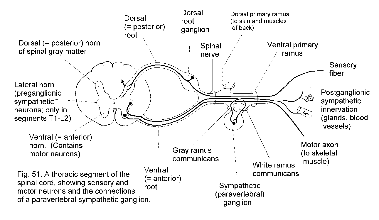
Also:
http://images.google.com/imgres?imgurl=http://upload.wikimedia.org/wikipedia/commons/thumb/e/ee/Gray799.svg/250px-Gray799.svg.png&imgrefurl=http://en.wikipedia.org/wiki/Gray_ramus_communicans&usg=__qUXCvvpEp5jq-MU-9iTXPLFa0f0=&h=222&w=250&sz=31&hl=en&start=8&sig2=C5MPFfY1uQFQtlrMe7Nwww&um=1&tbnid=9BHh2mc5_tWNJM:&tbnh=99&tbnw=111&prev=/images%3Fq%3Dgray%2Brami%2Bcommunicans%2Bmodel%26hl%3Den%26um%3D1&ei=AFXqSortMMSztgfR-9w_

Spinal Cord Cross Section:
Gray horns (anterior, lateral posterior), white columns (anterior, lateral, posterior), central canal
http://nhscience.lonestar.edu/bioL/ap1model/labeled/spinalcord3.jpg

http://bioweb.uwlax.edu/Aplab/Table_of_Contents/Lab_07/Spinal_Model_1/spinal_model_1.html
http://bioweb.uwlax.edu/Aplab/Table_of_Contents/Lab_07/Spinal_Model_2/spinal_model_2.html
Nerve and Nerve Plexi / Cranial Nerves:
Using appropriate models, be able to identify all 12 pairs of cranial nerves as well as the primary nerves of the cervical plexus, brachial plexus, lumbar plexus, and sacral plexus. Be sure to review the function of each.
http://nhscience.lonestar.edu/bioL/ap1model/labeled/spinalcord.jpg

The secret to remembering these is to know that the are numbered in order from rostral to caudal end, starting with the Olfactory nerve. From there, you can count off the nerves and use the mneumonic "OLd OPie OCcassionally TROmps TRIGonometry And Feels VEry GLOomy, VAGUe And HYPOactive."
http://content.answers.com/main/content/img/oxford/Oxford_Body/019852403x.cranial-nerves.1.jpg

http://academic.kellogg.edu/herbrandsonc/bio201_McKinley/f15-24l_cranial_nerves_c.jpg

Quiz yourself on Cranial Nerves!
http://www.dontbeasalmon.net/elearning/term3spotters/docs/qa/CN.html
Sympathetic Efferent Pathway Models:
Paravertebral sympathetic ganglia, Prevertebral sympathetic ganglia
The paravertebral sympathetic ganglia are also known as the sympathetic trunk ganglia or chain ganglia. The prevertebral ganglia are located further away from the sympathetic trunk (more distal?), and are also known in our text as collateral ganglia.
http://www.ncbi.nlm.nih.gov/bookshelf/br.fcgi?book=neurosci&part=A1391&rendertype=figure&id=A1393
Human Brain Model:
Cerebrum:
Cerebral hemispheres (left and right), transverse fissure, longitudinal fissure, lateral sulcus, central sulcus, parieto-occipital sulcus, precentral gyrus, postcentral gyrus, frontal lobe, parietal lobe, temporal lobe, occipital lobe, insula, basal nuclei, corpus callosum, septum pellucidum, fornix, cortex, internal capsule, olfactory bulbs, olfactory tracts, optic nerves, optic chiasma, optic tracts, roots of nerves III – XII, lateral ventricles, choroid plexus
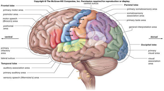
http://lh4.ggpht.com/_lRGislJejZ0/SRp7sb6QjHI/AAAAAAAAB8c/xeGXGZP0LUY/image20_thumb4.png?imgmax=800
http://mindsci-clinic.com/cerebral_lobes_and_insula.htm
Quiz yourself on the internal structures:
http://www.dontbeasalmon.net/elearning/term3spotters/docs/qa/index.html
Diencephalon:
Pituitary gland, mammillary bodies, thalamus, massa intermedia, hypothalamus, pineal body, interventricular foramen, third ventricle, choroid plexus, infundibulum

http://academic.kellogg.edu/herbrandsonc/bio201_McKinley/f15-15_diencephalon_c.jpg
http://academic.kellogg.edu/herbrandsonc/bio201_McKinley/f15-17_hypothalamus_c.jpg

Cerebellum:
Vermis, cortex, arbor vitae
http://academic.kellogg.edu/herbrandsonc/bio201_McKinley/f15-22a_cerebellum_mids_c.jpg

http://anatomia.wum.edu.pl/atlas/image_08e.htm
Brain Stem:
Cerebral aqueduct, fourth ventricle, median lateral aperatures, choroid plexus, midbrain, corpora quadrigemina, superior colliculi, inferior colliculi, cerebral penduncles, pons, middle cerebellar peduncles, pyramids & medulla oblongata
http://academic.kellogg.edu/herbrandsonc/bio201_McKinley/f15-20a_pons_longitudin_c.jpg

http://www.clinic-clinic.com/clncl-mdcne/nrsrgry/Dx/CSF%20circulation.htm
Meninges:
Dura mater (periosteal meningeal layer), arachnoid mater, subarachnoid space, pia mater, tentorium cerebelli, falx cerebri
If you scroll down a bit on this page to the section labeled "The Dynamic Shifting of Tensions in the Dura Mater", you will see a good illustration and explanation of the tentorium cerebelli and the falx cerebri.
http://www.osteodoc.com/sutherland.htm
Great model showing everything but the tentorium cerebelli:
http://academic.kellogg.edu/herbrandsonc/bio201_McKinley/f15-4_cranial_meninges_c.jpg

Sheep Brain Dissection:
Cerebrum, cerebral hemispheres (left and right), cerebellum, midbrain, pons, medulla oblongata, olfactory bulbs, olfactory tracts, longitudinal fissure, optic tracts, optic chiasma, lateral ventricles, third ventricle, fourth ventricle, corpus callosum, thalamus, hypothalamus, and pineal body
http://www.hometrainingtools.com/article.asp?ai=1316&bhcd2=1256927391
Really good quiz to test your knowledge:
http://www.gwc.maricopa.edu/class/bio201/brain/brshpx.htm
http://www.gwc.maricopa.edu/class/bio201/brain/brshpx2.htm
http://www.gwc.maricopa.edu/class/bio201/brain/brnshp5.htm
Brain Slides:
Pyramidal cells(from cerebrum) Purkinje cells (from cerebellum)
Pyramidal Cells:
http://neuromedia.neurobio.ucla.edu/campbell/nervous/wp_images%5C198_pyramidal_cells.gif

Purkinje Cells:
http://education.vetmed.vt.edu/Curriculum/VM8054/Labs/Lab9/IMAGES/PURKINJE%20LAYER%20COMPOSITE.jpg

Human Eye Model:
Lacrimal gland, lacrimal puncta, conjunctiva (palpebral and ocular), conjunctival sac, extrinsic eye muscles (superior oblique,inferior oblique, superior rectus, inferior rectus, medial rectus, and lateral rectus), palpebrae, palpebral fissue, medial and lateral canthi, levator palpebrae superioris, eyelids, eyelashes, sclera, cornea, scleral venous sinus, choroid, iris, pupil, ciliary body, ciliary muscles, ciliary processes, suspensory ligaments, lens, ora serrata, macula lutea, retina, optic disc, fovea centralis, vitreous humor, aqueous humor, anterior chamber, posterior chamber
http://media-2.web.britannica.com/eb-media/47/63347-004-610F94B5.gif

http://media-2.web.britannica.com/eb-media/30/91430-004-828719A3.jpg

Cow Eye Dissection:
Sclera, cornea, choroid, ciliary body, aqueous humor, vitreous humor, iris, pupil, optic nerve, lens, retina, & optic disc
http://district.bluegrass.kctcs.edu/rmccane0001/shared_files/bio137website/BIO137/137Lab10/Lab10CowEyeLabeled.html
Retina Slide:
Ganglion cell layer, bipolar cell layer, rods, cones, and pigmented layer
https://blogger.googleusercontent.com/img/b/R29vZ2xl/AVvXsEgMXEB_lH2oWP9sYDiapNviAtCARoO2mtnmWJ7IhTQa6wXqccTUMzuSnmi8wAELJK9ct_dew9g7_wb9UfeaQpeAXoO4elzBICg2Dpcr0XxH3cDjDJ-SuGm_cB7WGzqtdm1L6XR8TKcPkKXu/s1600-h/100_1965.jpg

Human Ear Model:
Outer ear, pinna, external auditory canal, tympanic membrane, middle ear, malleus, incus, stapes, eustachian tube, round window, oval window, inner ear, cochlea, cochlear duct, organ of Corti (basilar membrane, tectorial membrane, and hair cells), vestibule, vestibulocochlear nerve (including vestibular branch and cochlear branch), semicircular canals, semicircular ducts with ampulla, utricle, and saccule
http://www.infj.ulst.ac.uk/~pnic/HumanEar/Andy
http://mail.pittsfield.net/teachersites/Whelihan_Kathleen/0147FF93-000F6E5A.2/The-Human-Ear.gif

http://www.seanpalaciosmd.com/Vertigo.html

Cochlear Cross-Section Model:
Scala vestibule, vestibular membrane, cochlear duct, organ of Corti, tectorial membrane, basilar membrane, scala tympani, endolymph, perilymph, spiral lamina, and spiral ganglion
http://www.hearingcentral.com/images/inner_ear.gif

http://media-2.web.britannica.com/eb-media/01/14301-004-4B6F34DA.gif

http://www.oto-hns.northwestern.edu/Auditory%20Research%20Laboratory/tutorials.html
Cochlear Slide:
Scala vestibule, vestibular membrane, cochlear duct, basilar membrane, scala tympani, and organ of Corti
http://biology.clc.uc.edu/fankhauser/Labs/Anatomy_&_Physiology/A&P202/Special_Senses/Ear/cochlea_loop_P2171519labeled.jpg

https://blogger.googleusercontent.com/img/b/R29vZ2xl/AVvXsEiMun2fzlRyu-9J4oNsHYjhgvwha2-dsWiCVQTYAtuncatra2V4CkYFR_A3wYQ8mRmH8KSJURpiusGsqqFUdsiIXs8uYSkbUWb-6xUaM4imBbv9WJQDgldZ711gxUWqvc0ROosTaYDMwfeR/s1600-h/100_1961.jpg

Olfactory Epithelium Slide:
Olfactory cells and Cilia
https://blogger.googleusercontent.com/img/b/R29vZ2xl/AVvXsEio4tgSUNNXAgaD37FofvldovpdCM16xoCgXkfWJZg_KqK1JxzEct7gM2euf2MyLrsR_nBF9evvvWJP49cYK0Sq2Y3zX_ek97J38Mak9ruvXHOa3SwuecIVwwA18BR6UGoQE7nf6mq0D4_q/s1600-h/100_1962.jpg

Tongue Slide:
Papillae and Taste buds
https://blogger.googleusercontent.com/img/b/R29vZ2xl/AVvXsEhXDg0xB_Wh2nazE-pvfTJD5qCx4TZJksAwRL0Nj2WPctIbChJ71tApg1nXqcCSNk1nQY1SMzBv5WK7HcbSwtiY7gkZuVHombAedR2cILveJgIzE8jRZJDYuPDjKihLeghOqUm5femcJxEV/s1600-h/100_1963.jpg

Endocrine System Lab:
Full Endocrine System Model (minus the subsections):
Hypothalamus, pituitary (adenohypophysis and neurophypophysis), thyroid, parathyroid, thymus, pancreas, adrenal gland (adrenal cortex and adrenal medulla), ovaries, testes, and pineal gland
Scroll down a bit on the page to the endocrine system portion...
http://district.bluegrass.kctcs.edu/rmccane0001/shared_files/bio137website/BIO137/137Lab1/Lab1Organs.html
pituitary slide showing adenohypophysis and neurohypophysis:
http://www.siumed.edu/~dking2/erg/enguide.htm#pituit
adrenal gland with information on adrenal cortex, adrenal medula, and the various regions of cell histology:
Sweet little point and click web app that shows the adrenal glad, it's gross anatomy, and histology from Boston University:
http://www.bu.edu/histology/p/14501ooa.htm
Endocrine System Slides:
Pituitary slide:
Identify the following histological features of the pituitary:
Adenohypophysis and neurohypophysis
Thyroid slide:
Identify the following histological features of the thyroid:
Follicles, follicular cells, thyroglobulin, and parafollicular cells
Parathyroid slide:
Identify the following histological features of the parathyroid:
Chief cells
Adrenal gland slide:
Using a slide of the human adrenal gland, be able to distinguish between the following:
Adrenal cortex, zona glomerulosa, zona fasciculate, zona reticularis, adrenal medulla, chromaffin cells
Pancreas slide:
Using a slide of the human pancreas, be able to identify the Islets of Langerhans
For all slides (with the exception of the Parathyroid slide) check here:
http://district.bluegrass.kctcs.edu/shirley.whitescarver/BIO139Lab/BIO139/139Lab12/Lab12EndocrineSlides.html
For Parathyroid, check here:
http://instruction.cvhs.okstate.edu/histology/histologyreference/hrendo.htm
A Fun Pancreas Point and Click Web App from Boston University:
http://www.bu.edu/histology/p/10403ooa.htm
Subscribe to:
Posts (Atom)